Step by step
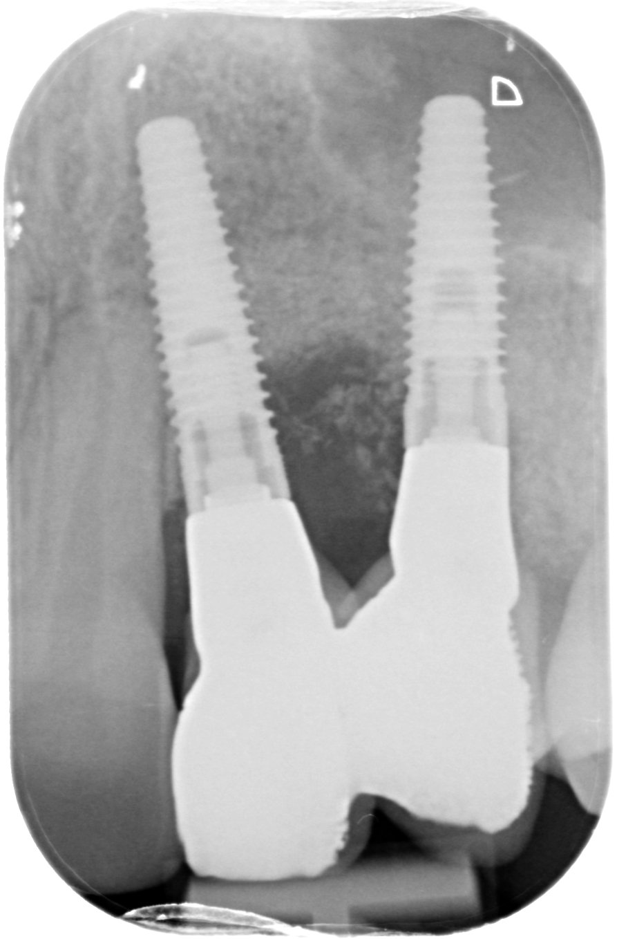
Figure 1.
Radiographic view of the failing implant-supported restoration at 22 and 23.
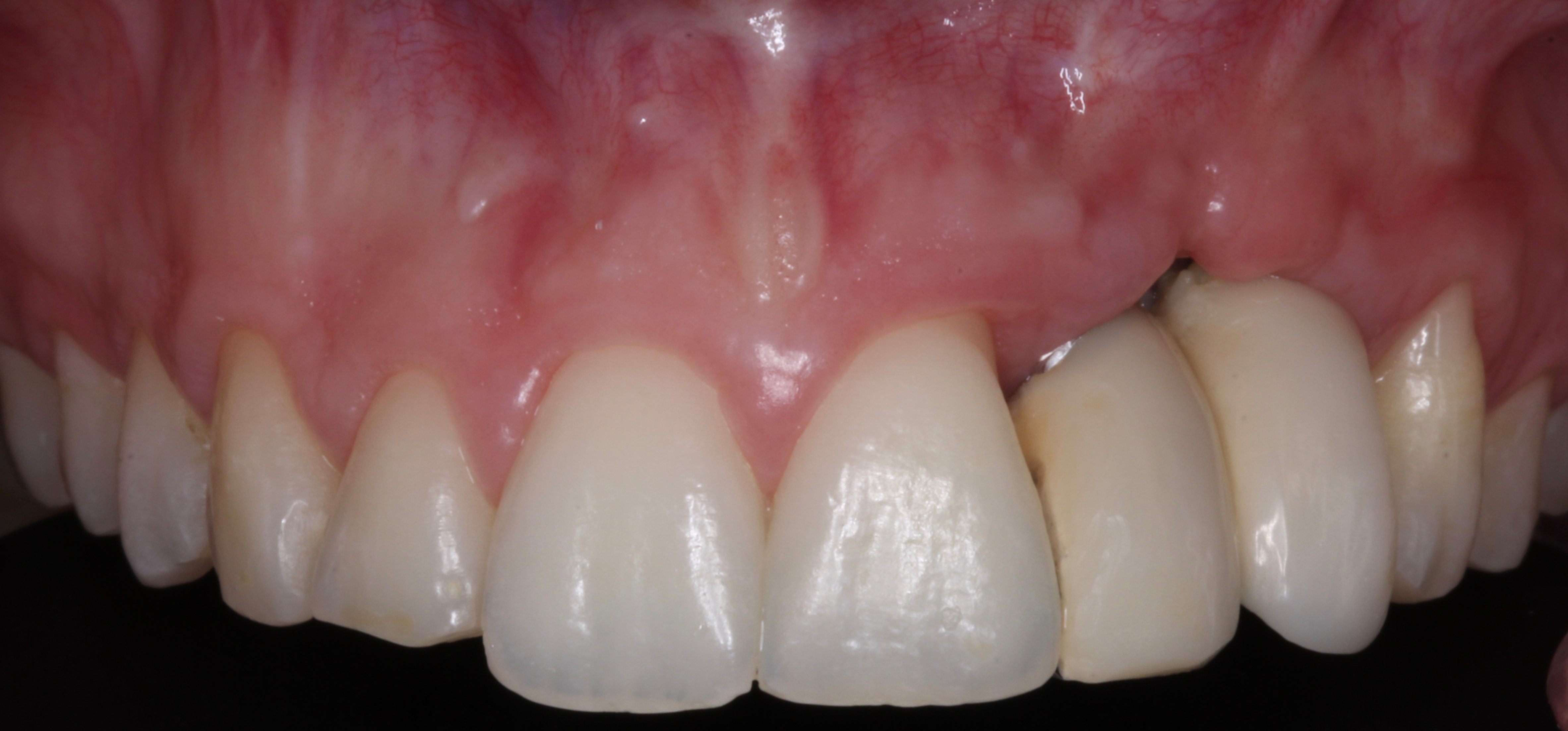
Figure 2.
Clinical view of the failing implant-supported restoration at 22 and 23.
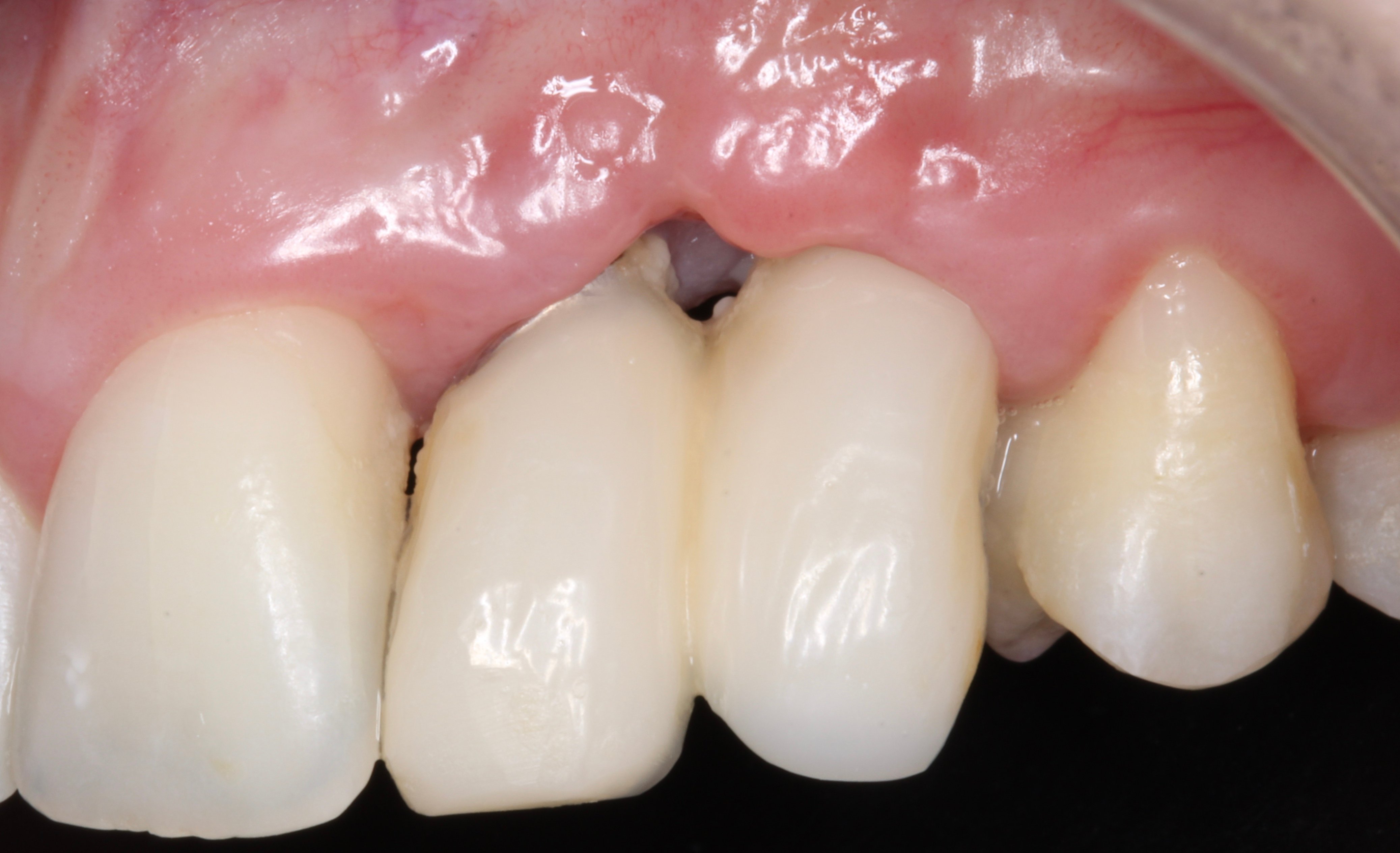
Figure 3.
Observe the papilla loss between them and the gingival recession at distal aspect of 21.
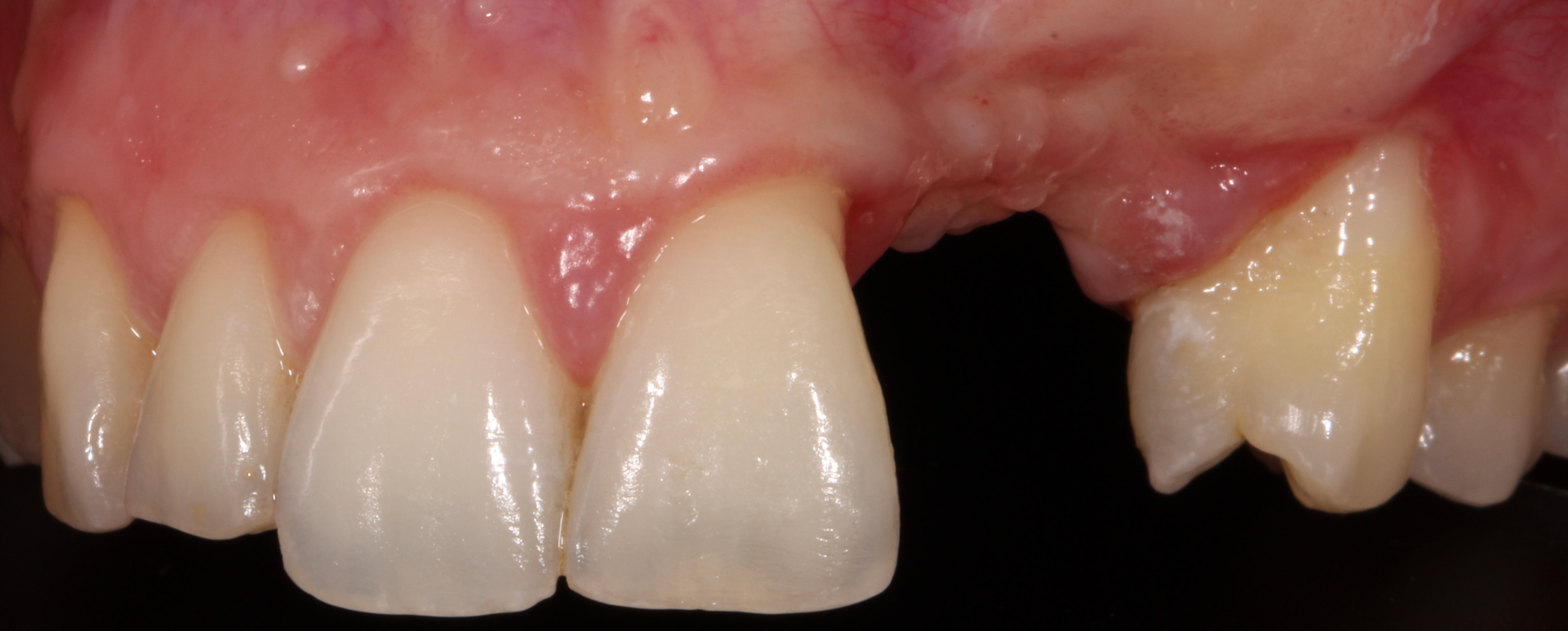
Figure 4.
Clinical view of the alveolar ridge 2 years after explantation. The patient was treated elsewhere and failed. Observe the increase of the gingival recession at distal aspect of 21 and the presence of scars.
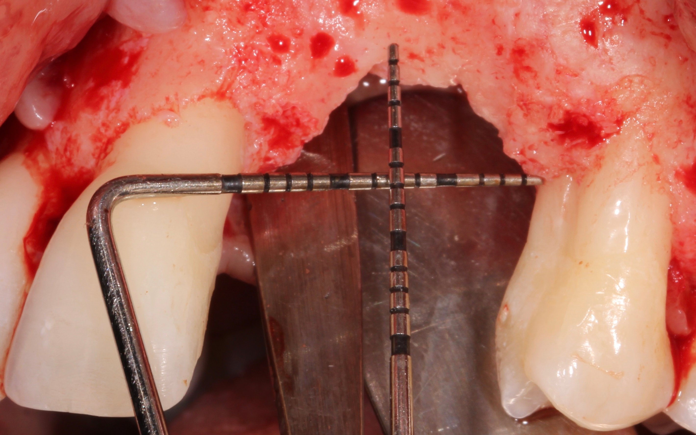
Figure 5.
Intrasurgical view of the 3D bone loss showing a 7 mm vertical defect.
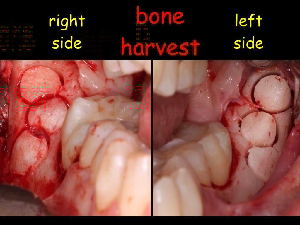
Figure 6.
Bone harvesting from both mandibular external oblique lines.
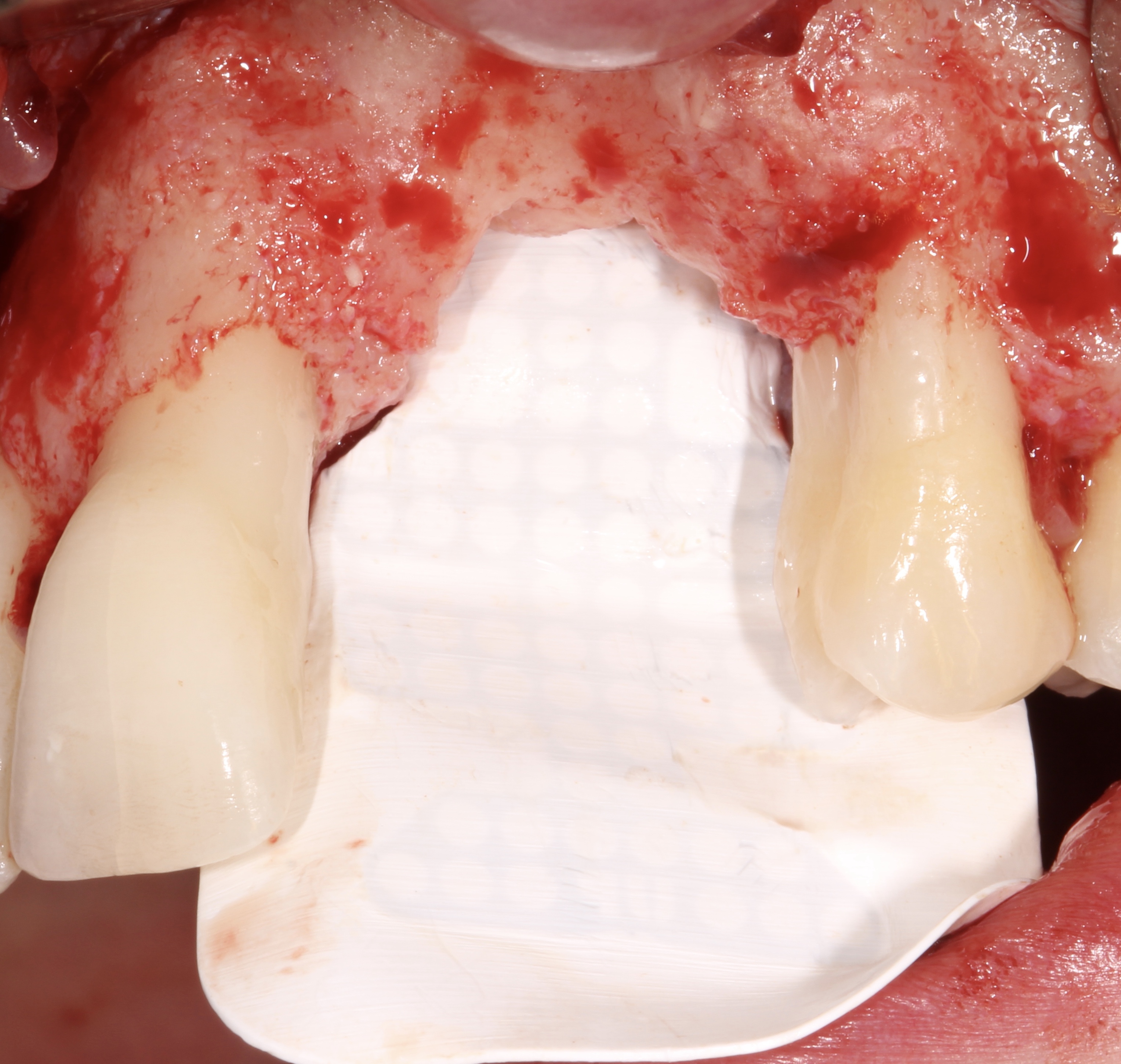
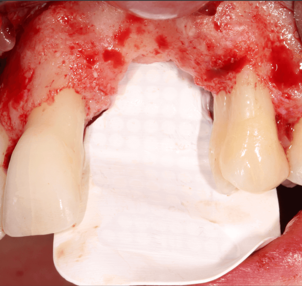
Figure 7.
Fixation of the NeoGen® membrane at palatal aspect.
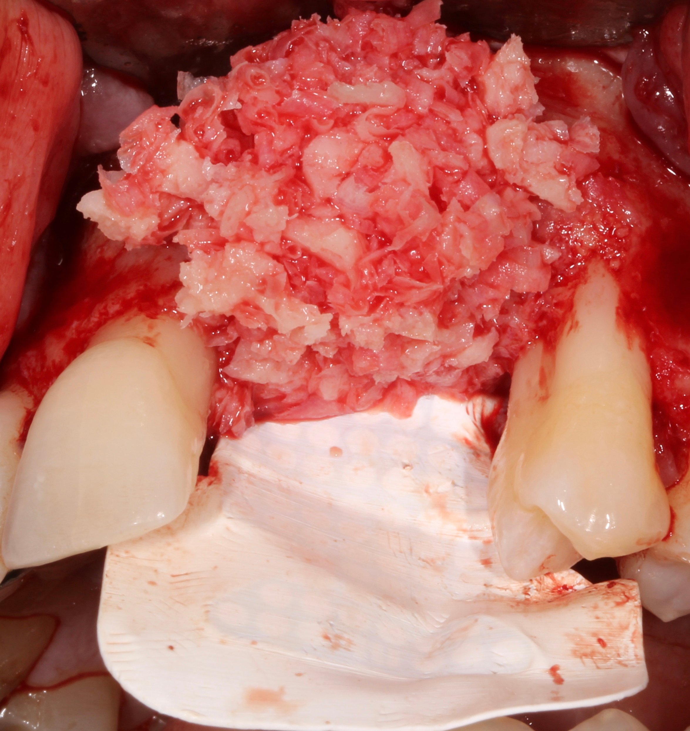
Figure 8.
Tension-free primary closure through correct management of the lingual and vestibular flaps and appropriate periosteal incisions, together with the application of simple horizontal mattress sutures.
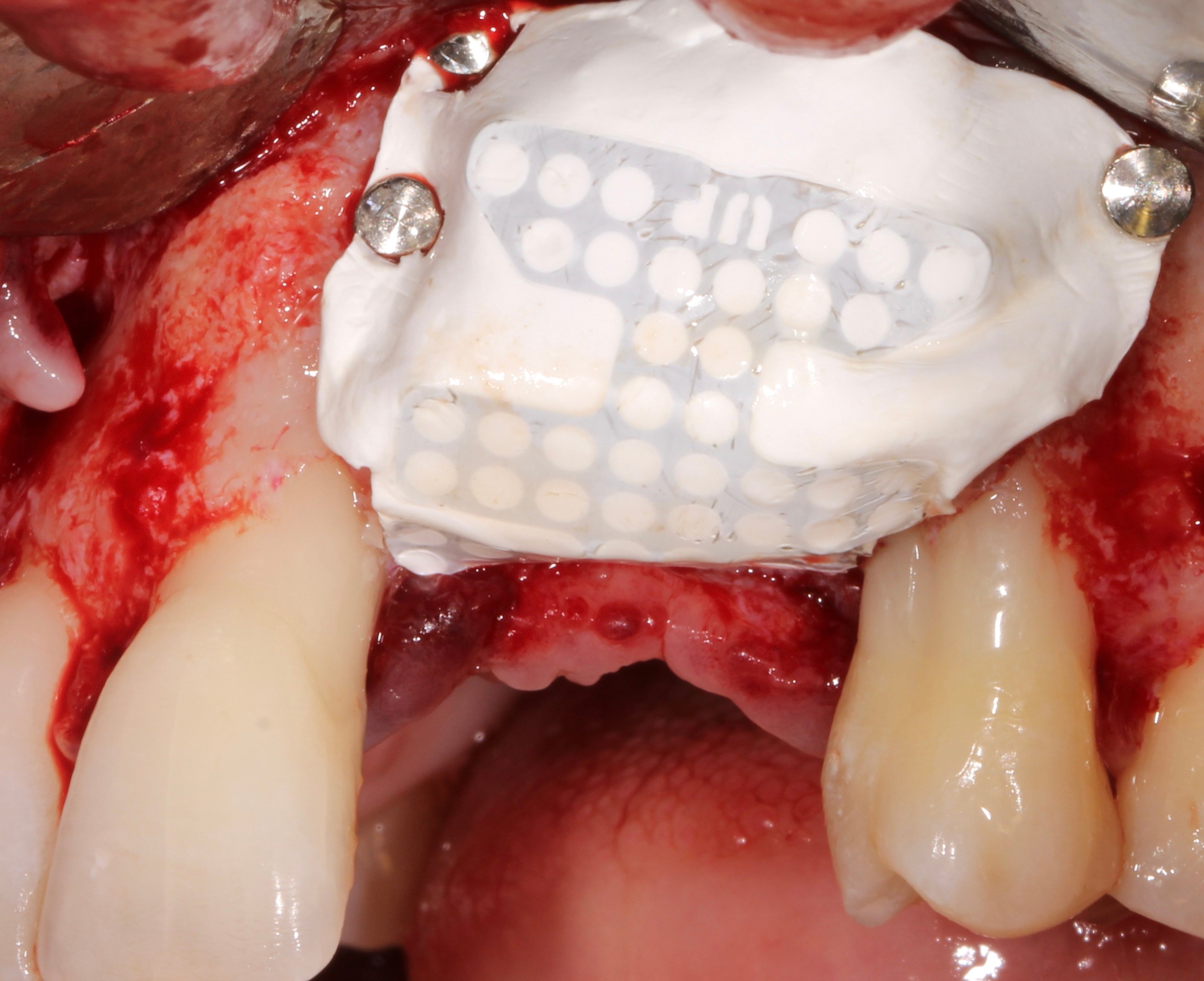
Figure 9.
Membrane fixed by Tacks. Observe that the coronal placement of the membrane is at the same level as the inter proximal bone peaks.
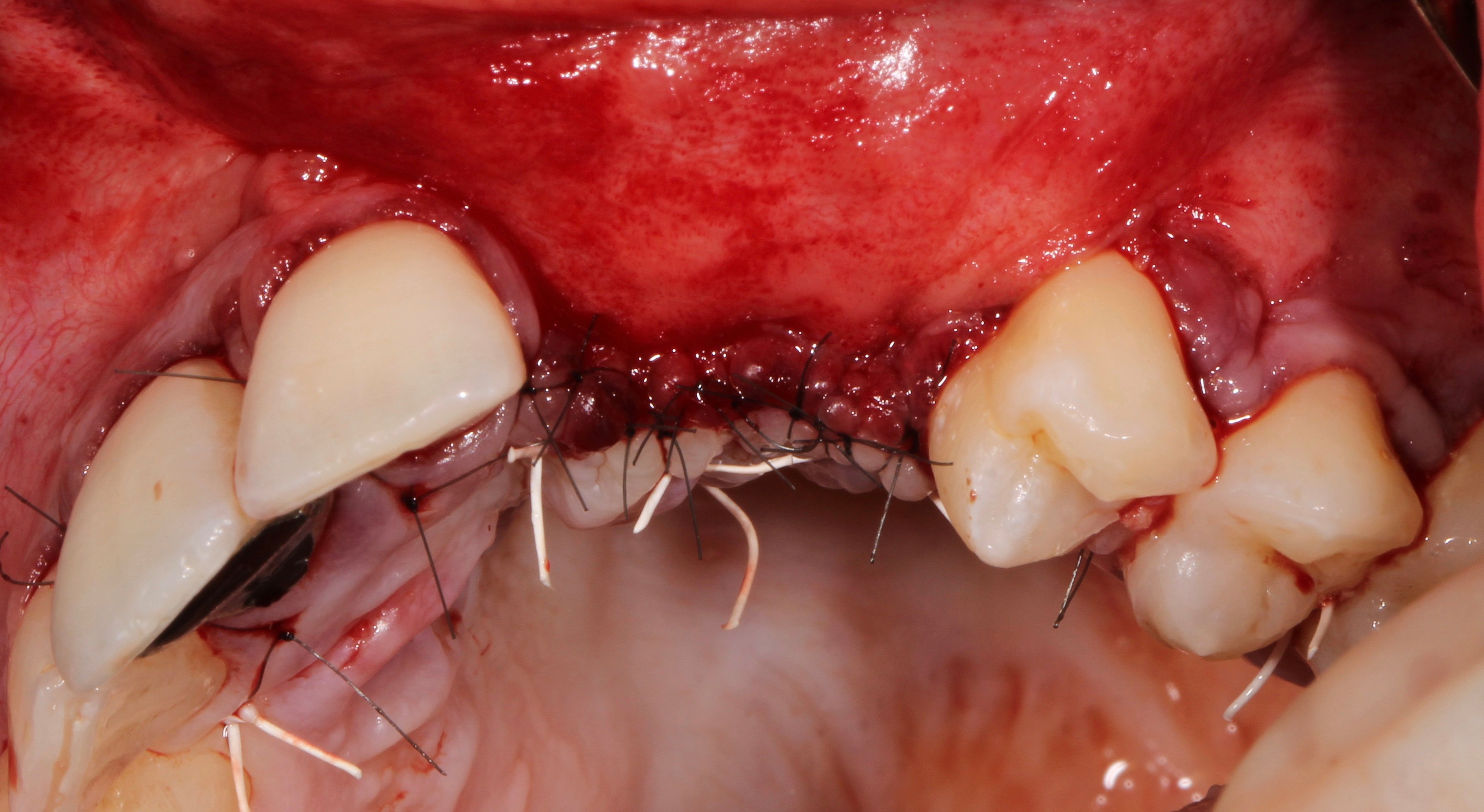
Figure 10.
Surgical suture closure.
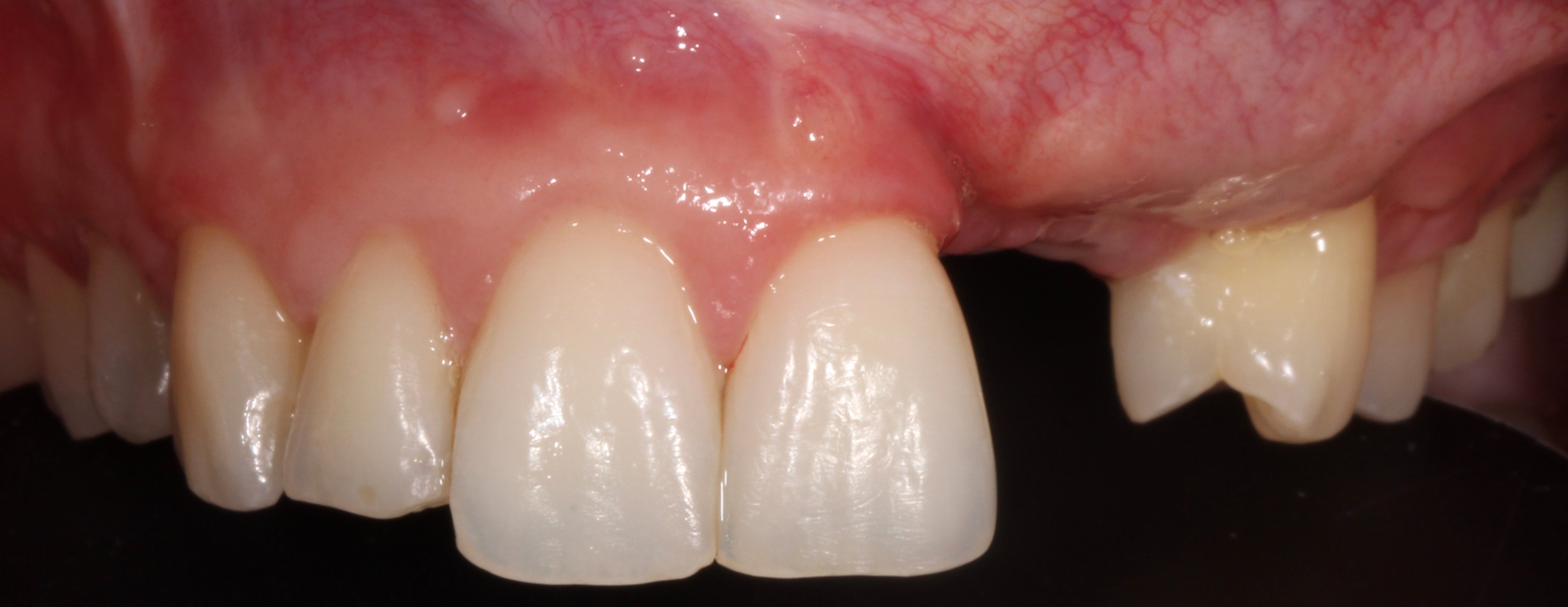
Figure 11.
Reentry 1 year after surgery.
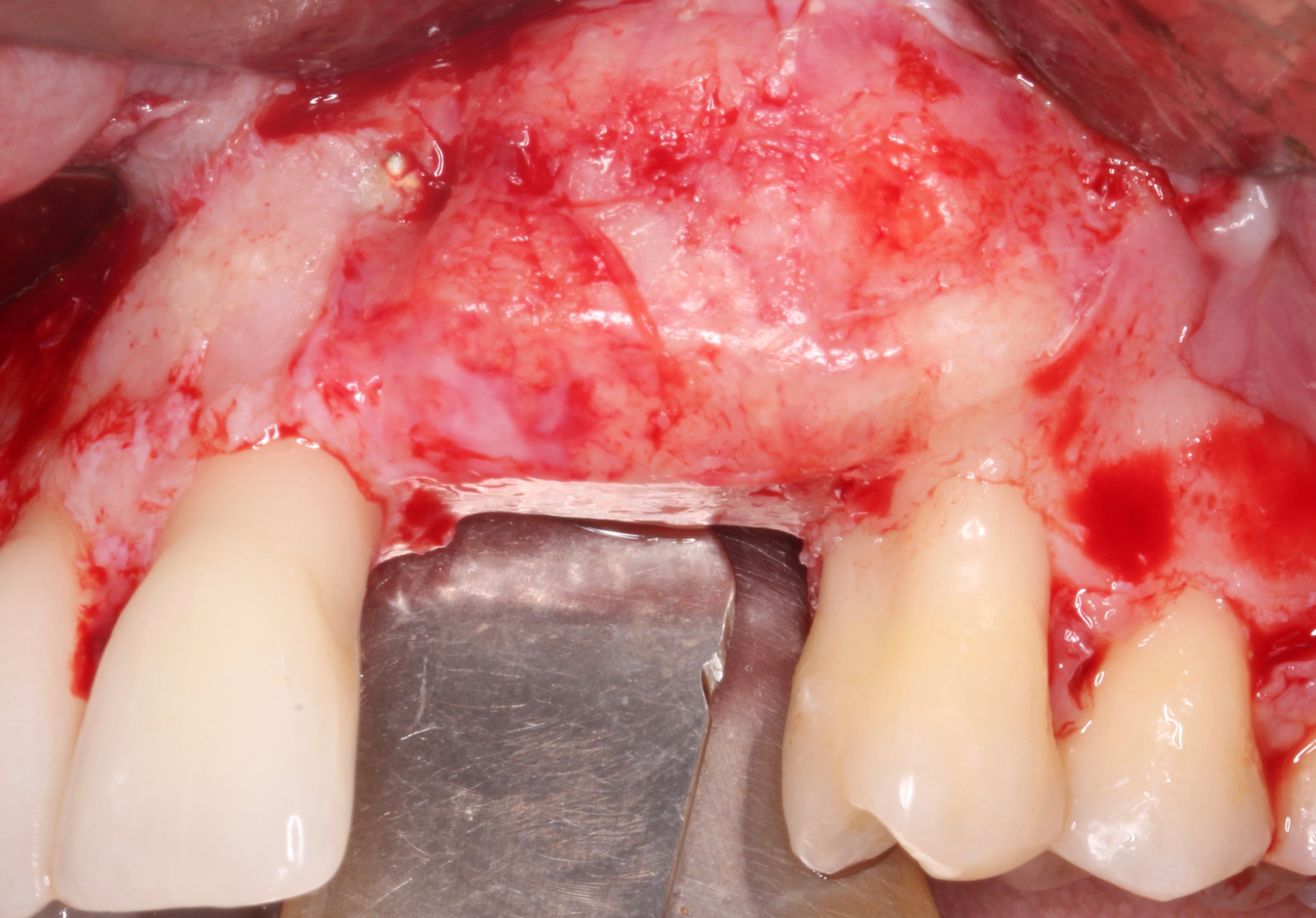
Figure 12.
Complete vertical bone regeneration
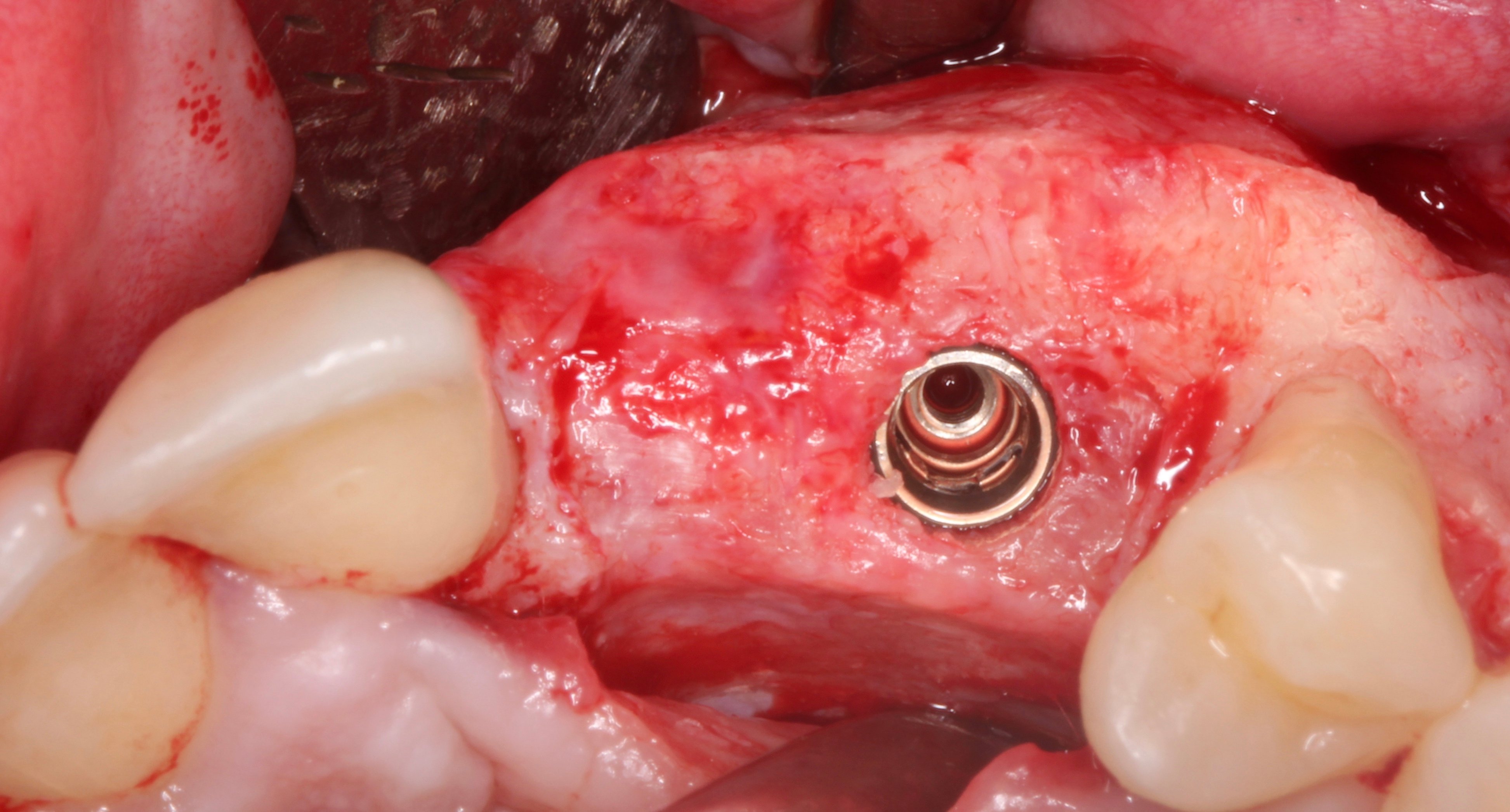
Figure 13.
Implant placed at 23 level to support 2 crowns (22 will be a cantilever).
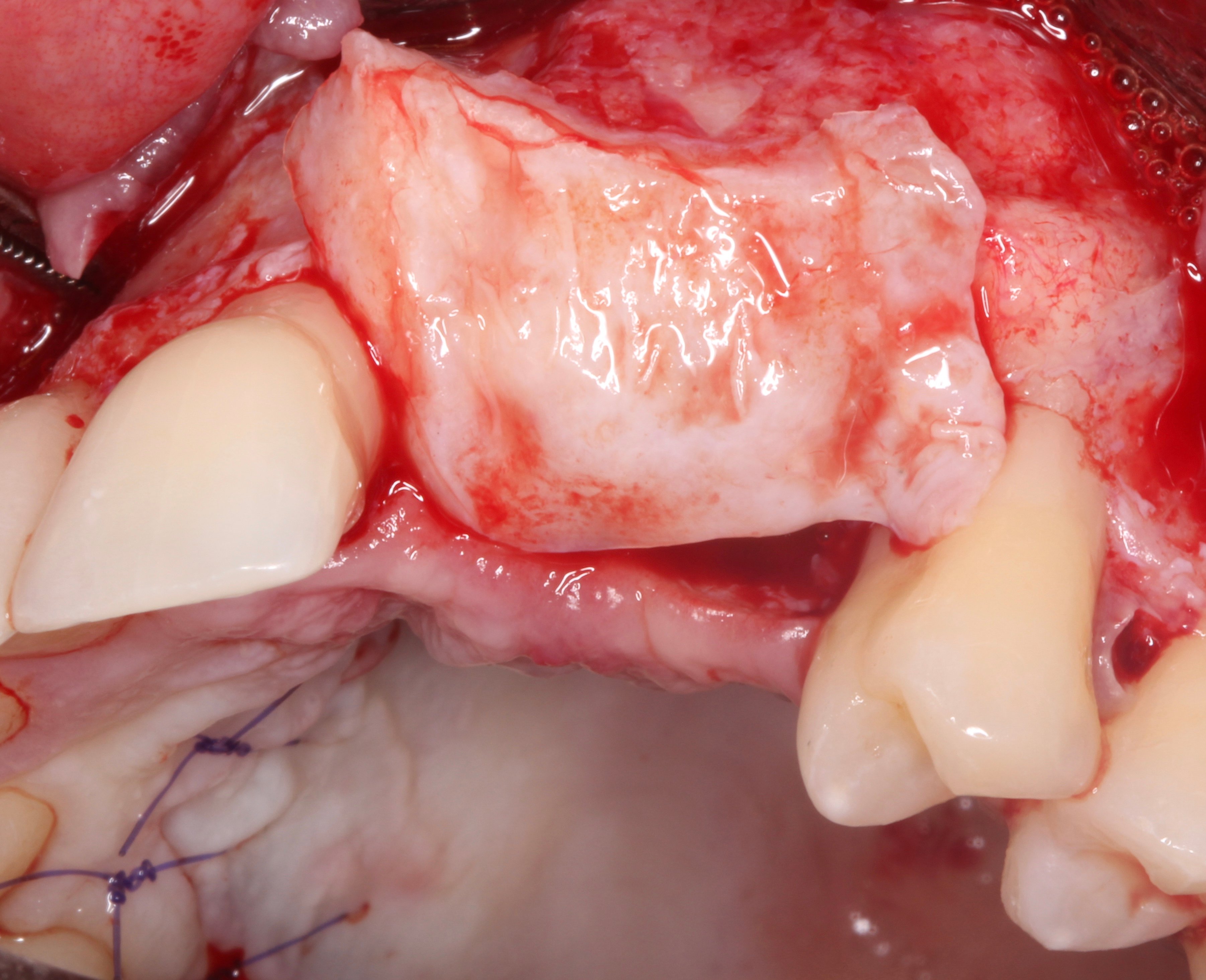
Figure 14.
Connective tissue graft folded and adapted to the distal aspect of 21 to reconstruct the papilla.
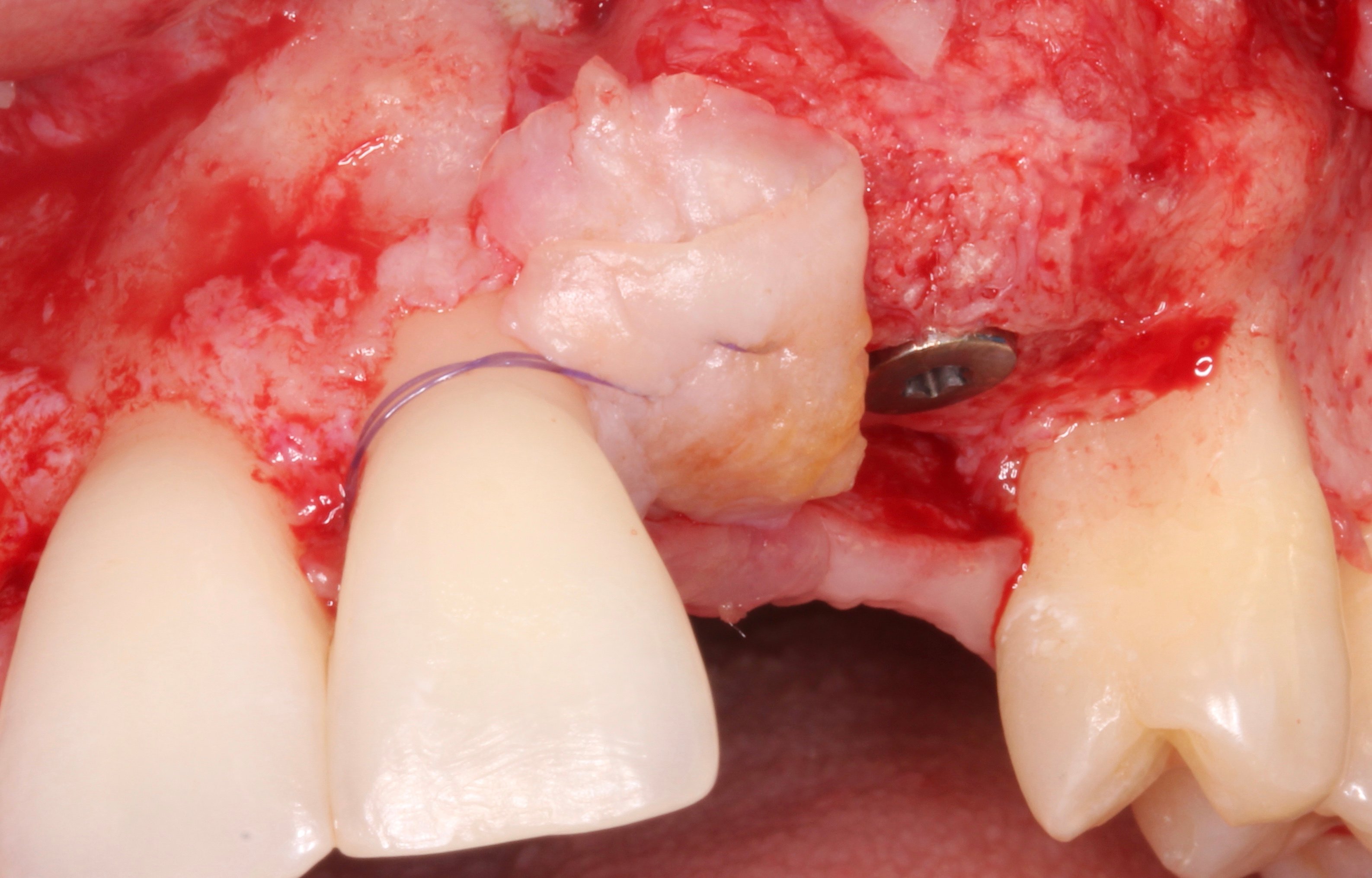
Figure 15.
Connective tissue graft folded and adapted to the distal aspect of 21 to reconstruct the papilla.
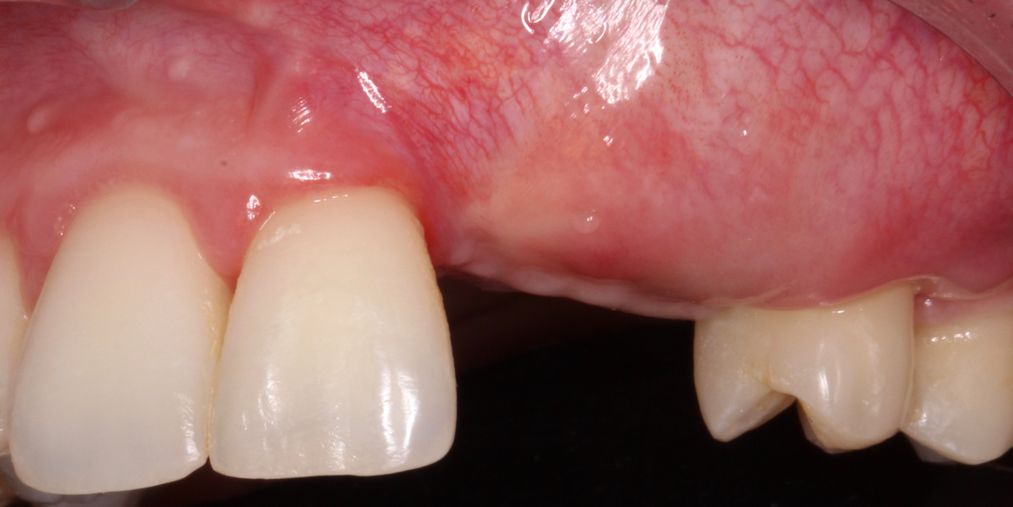
Figure 16.
Aspect 2 months later. The mucogingival line is displaced coronally and a surgery to reposition it is mandatory.
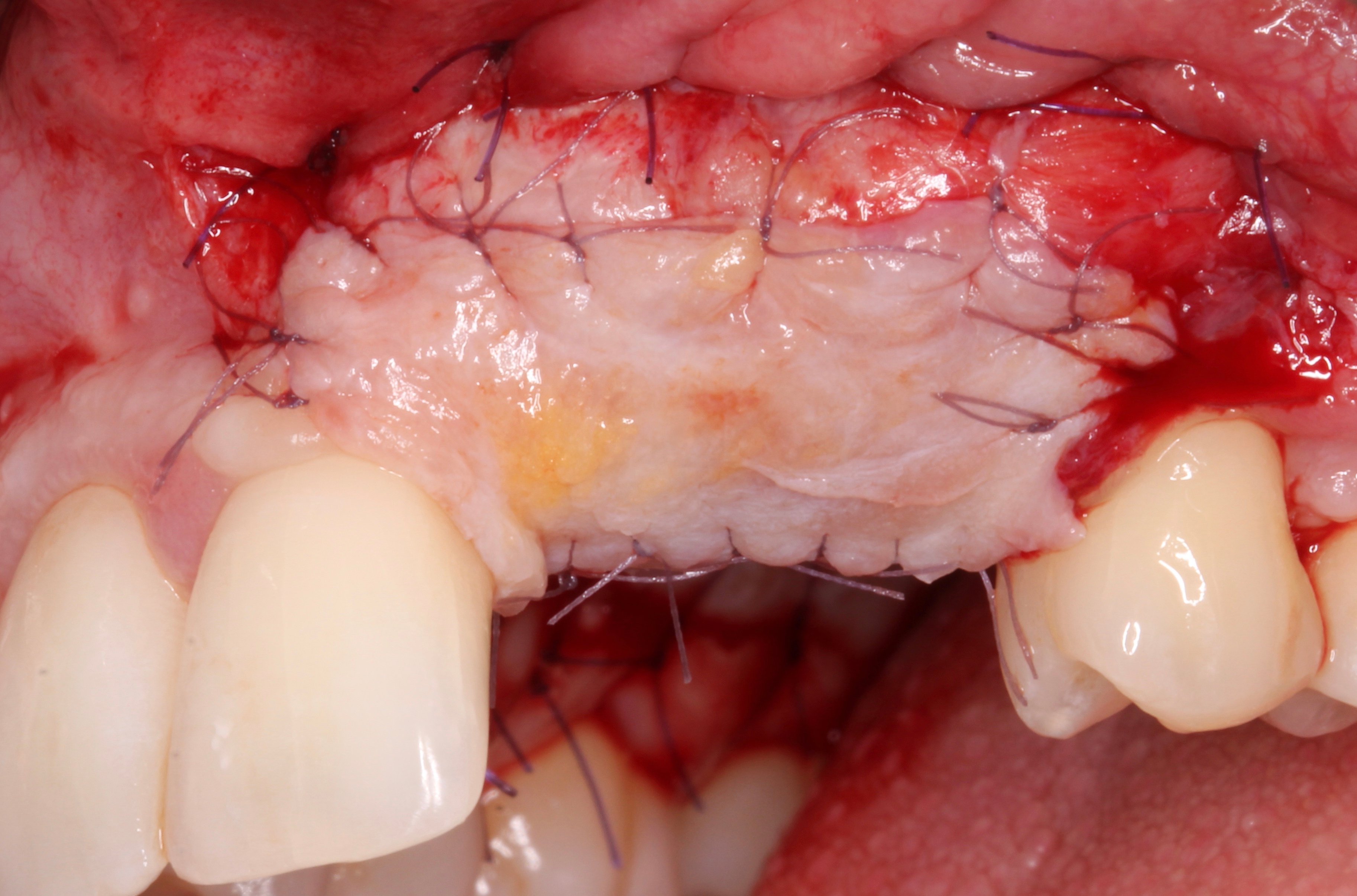
Figure 17.
Free Connective tissue graft to reposition the mucogingival line.
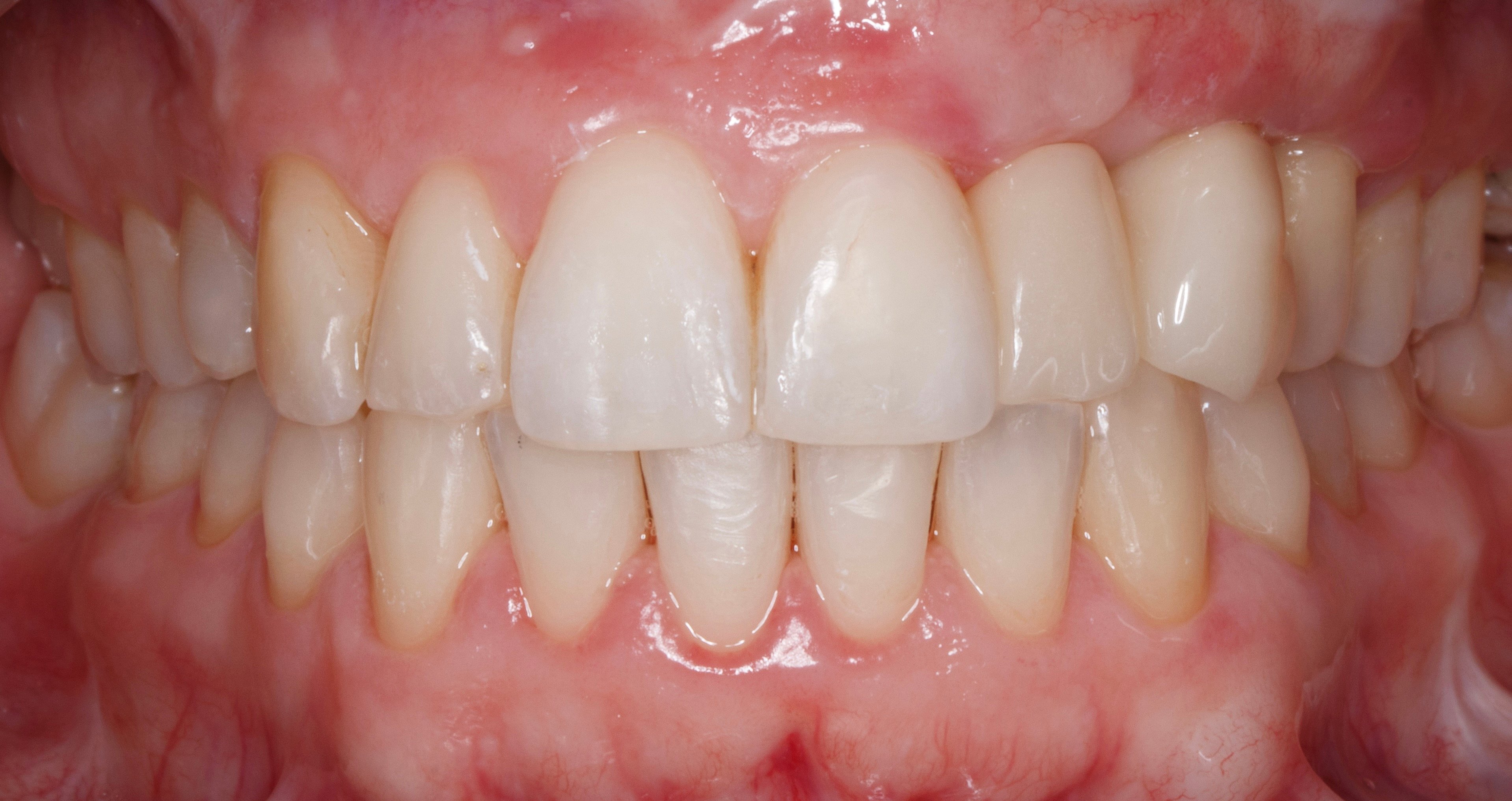
Figure 18.
Final prosthesis 6 years after loading.
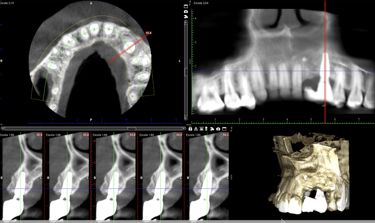
Figure 19.
Sagital view of the implant (CBCT). Observe the stability of the bone at vertical and horizontal aspect.
"After 28 years using PTFE membranes for GBR I strongly believe that hybrid (dense and expanded) NeoGen® membranes are the best since they have a perfect combination of malleability and resistance due to the soft, easy to shape but strong titanium mesh. I dominate other regenerative techniques but no doubt that my first choice for regeneration of 3D bone defects is NeoGen® membranes."
Dr. David González Spain 
Downloads