Step by step
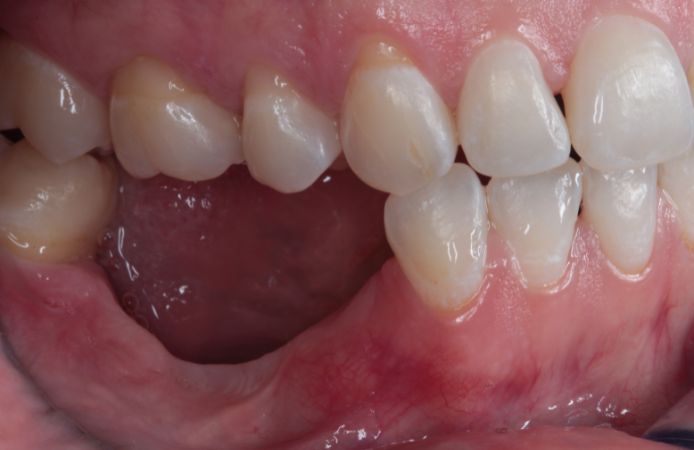
Figure 1.
Initial clinical situation. Severely resorbed posterior mandible following a previous failed attempt to insert dental implants with a split-crest technique.
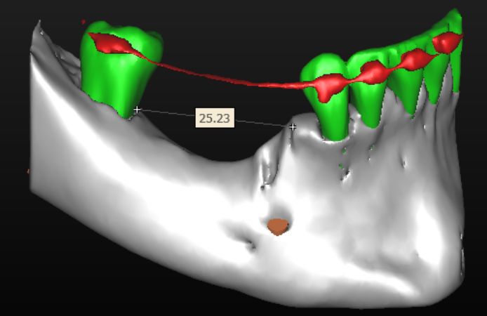
Figure 2.
CBCT evaluation showing a major bone defect in the posterior area of the mandible, with a length of 25 mm and a height of about 10 mm.
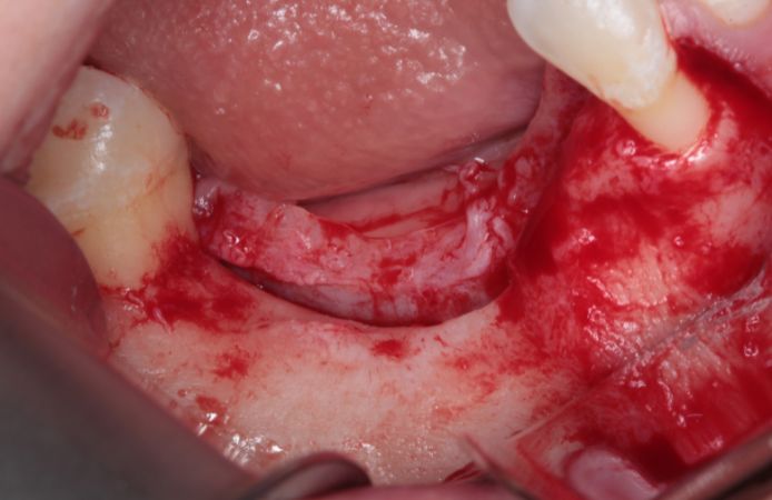
Figure 3.
Bone defect visible after full thickness flap elevation. A mid-crestal incision was performed with intrasulcular incisions. Vertical incisions were performed one tooth away from the defect.
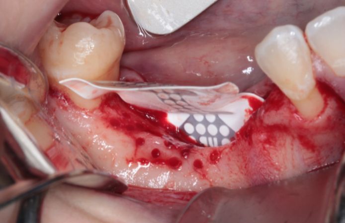
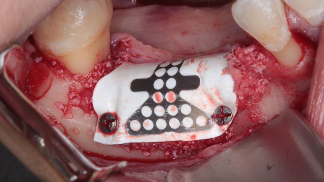
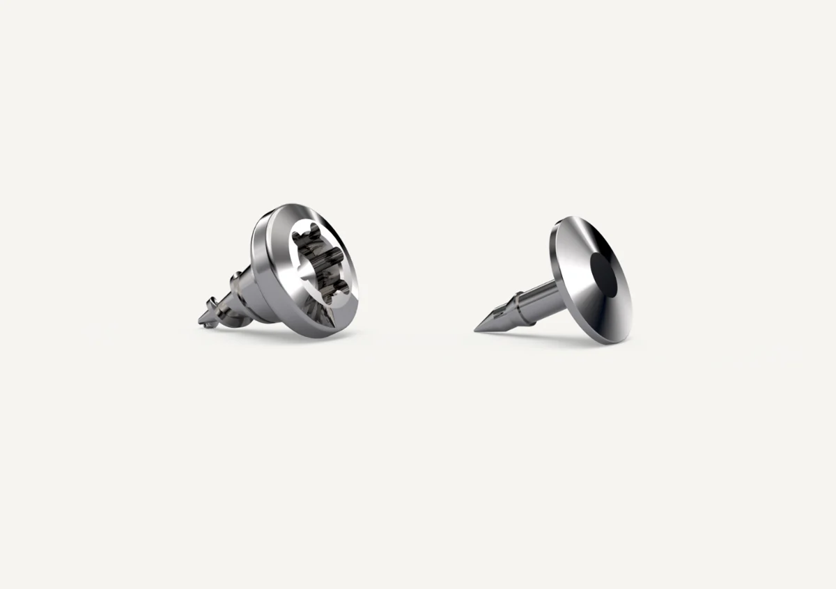
Figure 4.
The site was prepared with bone perforation. A NeoGen Ti-Reinforced PTFE Membrane was inserted and fixed lingually with two screws. The titanium-reinforced membrane provides structural support for space maintenance and undisturbed bone regeneration.
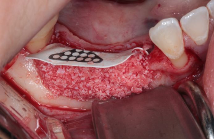
Figure 5.
Bone augmentation. Positioning of bone graft (50% autologous bone and 50% deproteinized xenograft) to support site augmentation.
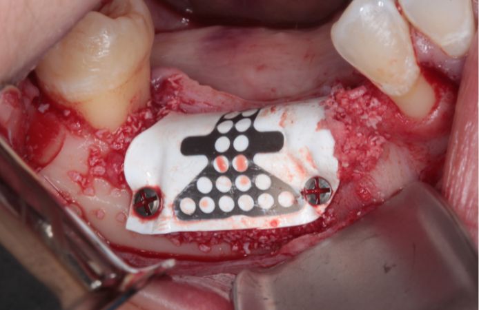
Figure 6.
The membrane is fixed buccally with two screws under tension to ensure good stability of the augmentation site.
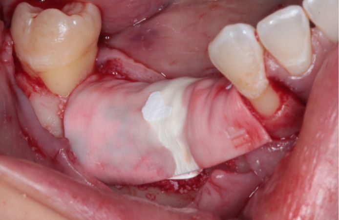
Figure 7.
Full coverage of bone graft particulates. A collagen membrane was used to cover the bone particulate close to the adjacent teeth that were not covered by the PTFE membrane.
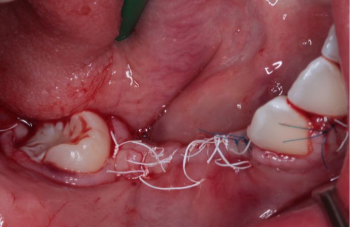
Figure 8.
Tension-free primary closure. Periosteal flap releasing incisions in order to achieve a tension-free and complete primary closure of the site.
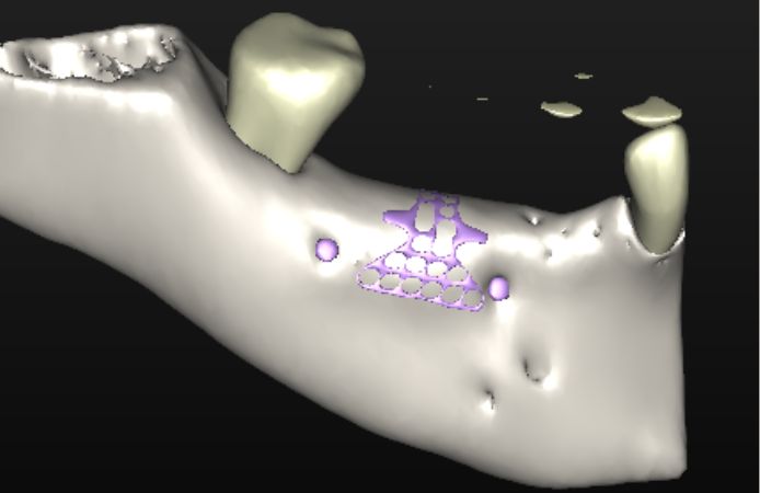
Figure 9.
CBCT after nine months of healing. Note the level of regenerated bone up to the NeoGen Ti-Reinforced PTFE Membrane.
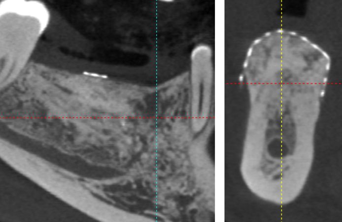
Figure 10.
CBCT evaluation after nine months of healing, note the vertical and horizontal bone regeneration, filling the whole volume under the NeoGen Ti-Reinforced PTFE Membrane.
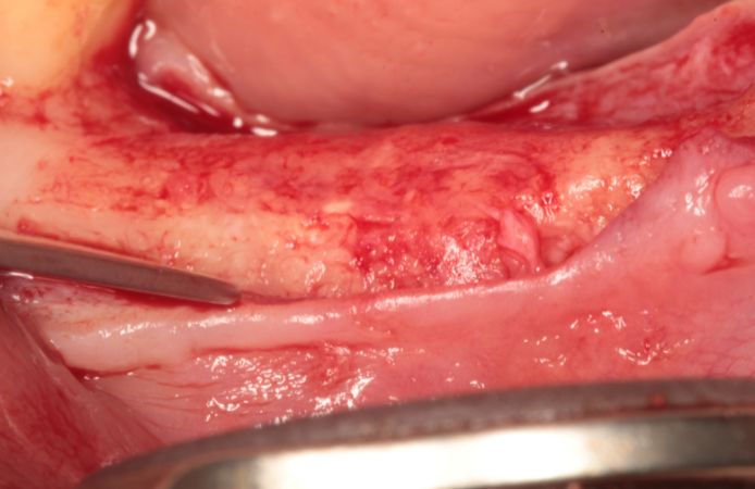
Figure 11.
Re-entry nine months after augmentation, after removal of the membrane. Note the level and width of regenerated bone. The newly formed bone is highly vascularized as indicated by abundant bleeding during site preparation for implant placement.
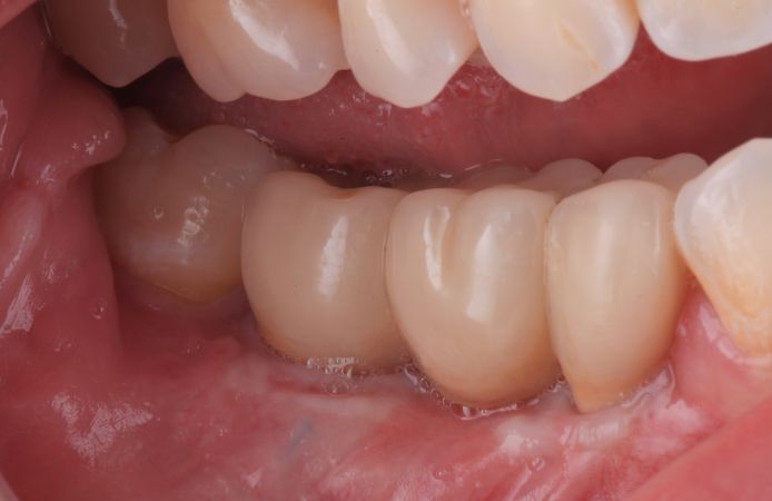
Figure 12.
Final restoration delivered six months after implant placement. The tissue levels are stable around the restoration with a satisfactory esthetic result.
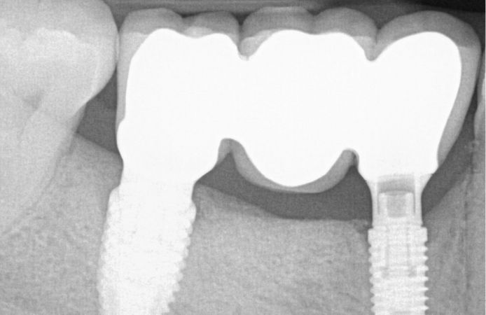
Figure 13.
Radiograph six months after implant placement with final restoration. The bone level is stable at implant level.
“The dual PTFE layer of the NeoGen membranes allows for optimal soft and hard tissue integration. The clinical handling is excellent, and its use allows for regenerating large three-dimensional bone volumes”
Isabella Rocchietta, DDS, MSc, Specialist in Periodontics, Italy 

Downloads
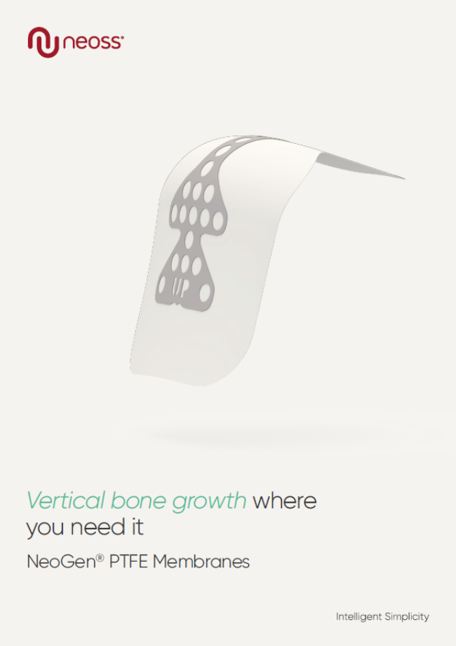
NeoGen PTFE Membranes
.png)